
High-Risk Maternal Care
Safeguarding Your Baby Through Autoimmune Challenges.
A maternal diagnosis of SLE (Lupus), Antiphospholipid Syndrome (APS), or Sjogren's requires vigilant fetal surveillance. Dr. Kunda Shahane provides the expert imaging needed to protect your baby's heart and monitor their growth trajectory.
Schedule Fetal AssessmentYour Antibodies, Your Baby's Health.
An autoimmune disease occurs when the body's immune system mistakenly attacks its own tissues. During pregnancy, specific maternal antibodies—such as Anti-Ro/SSA and Anti-La/SSB, commonly found in Lupus (SLE) and Sjogren's Syndrome—can cross the placenta and enter the baby's bloodstream.
Having an autoimmune condition does not mean a healthy pregnancy is impossible. In fact, with proper collaborative care between your Rheumatologist and a Fetal Medicine Specialist, the vast majority of mothers deliver beautiful, healthy babies. However, it mandates a highly specialized protocol of ultrasound and Doppler monitoring to detect early signs of fetal stress.
Conditions We Actively Screen For
Autoimmune conditions require targeted tracking of specific fetal risks.
Fetal Heart Block (Neonatal Lupus)
Maternal Anti-Ro/SSA antibodies can affect the electrical conduction system of the baby's heart, leading to a dangerously slow heart rate (Congenital Heart Block). Early detection via Fetal Echocardiography allows for potential medical intervention to prevent permanent damage.
Fetal Growth Restriction (FGR)
Autoimmune conditions, particularly Lupus, can cause inflammation in the blood vessels. This may compromise the placenta's ability to deliver adequate oxygen and nutrients, leading to a baby that is smaller than expected for their gestational age.
Pre-Eclampsia & Blood Clots
Mothers with Antiphospholipid Syndrome (APS) or Lupus have a higher risk of developing blood clots in the placenta and early-onset pre-eclampsia. We utilize precise Doppler scans of the uterine arteries to assess this risk early in the pregnancy.
Oligohydramnios
A reduction in amniotic fluid can occur if the placenta is not functioning optimally or if fetal kidney blood flow is restricted. We measure amniotic fluid index (AFI) rigorously during your third-trimester scans.
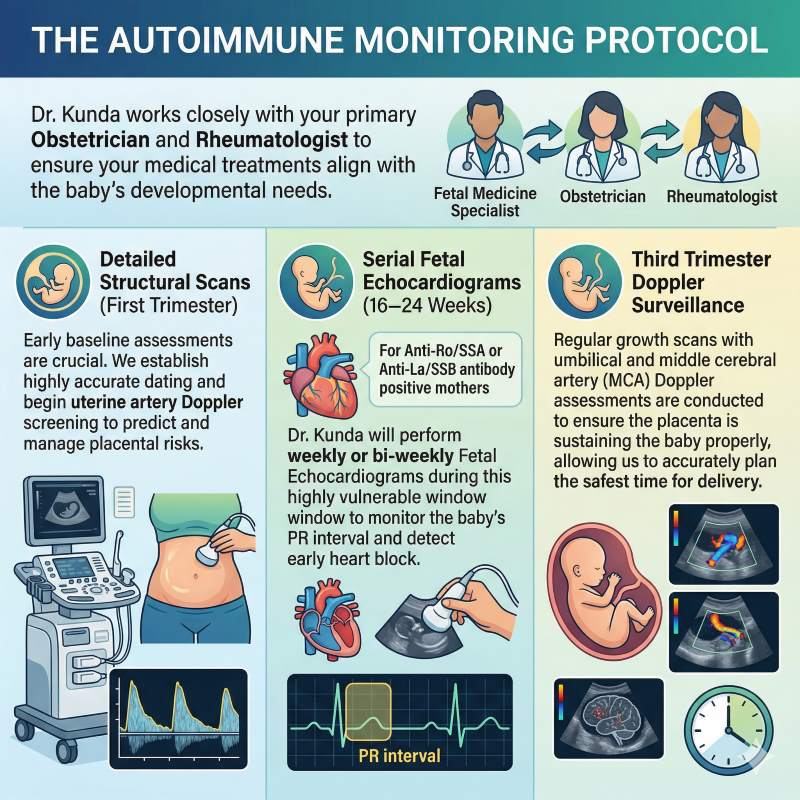
The Autoimmune Monitoring Protocol
Dr. Kunda works closely with your primary Obstetrician and Rheumatologist to ensure your medical treatments align with the baby's developmental needs.
Detailed Structural Scans (First Trimester)
Early baseline assessments are crucial. We establish highly accurate dating and begin uterine artery Doppler screening to predict and manage placental risks.
Serial Fetal Echocardiograms (16–24 Weeks)
If you test positive for Anti-Ro/SSA or Anti-La/SSB antibodies, Dr. Kunda will perform weekly or bi-weekly Fetal Echocardiograms during this highly vulnerable window to monitor the baby's PR interval and detect early heart block.
Third Trimester Doppler Surveillance
Regular growth scans with umbilical and middle cerebral artery (MCA) Doppler assessments are conducted to ensure the placenta is sustaining the baby properly, allowing us to accurately plan the safest time for delivery.