
Advanced Cardiac Imaging
A Closer Look at Your Baby's Heart.
A Fetal Echocardiogram is a highly specialized, non-invasive ultrasound dedicated entirely to mapping the structure, blood flow, and electrical rhythm of your baby’s heart. Dr. Kunda Shahane provides the expert screening needed for profound peace of mind.
Schedule Fetal EchoWhy Was I Referred for a Fetal Echo?
A referral for a Fetal Echo can immediately cause anxiety. It is incredibly important to know that most babies referred for this scan have perfectly healthy hearts. A standard 20-week anatomy scan checks the heart's basic four chambers, but a Fetal Echo is a microscopic, highly detailed audit of the cardiac walls, valves, and crossing vessels.
Because Congenital Heart Defects (CHDs) are the most common of all birth anomalies, Fetal Medicine Specialists perform this scan out of an abundance of caution. Many maternal conditions—like diabetes or undergoing IVF—slightly raise the statistical risk, making this scan a mandatory, protective step in your care plan.
Common Reasons for a Fetal Echo
You may be referred for this specialized scan based on maternal, fetal, or genetic factors.
Maternal Diabetes
Whether pre-existing (Type 1 or 2) or Gestational, fluctuating maternal blood sugar levels during the first trimester can directly impact how the baby's heart structures form, requiring a detailed structural audit.
IVF or ICSI Conception
Medical guidelines mandate a Fetal Echo for all pregnancies conceived through Assisted Reproductive Technology (ART). Studies show a slightly increased risk of cardiac anomalies in IVF and specifically ICSI pregnancies.
Autoimmune Conditions
Mothers with Lupus (SLE) or Sjogren’s Syndrome often carry Anti-Ro/SSA antibodies. These antibodies can cross the placenta and affect the baby's electrical conduction system, leading to a slow heart rate (heart block).
Family History of CHD
If you, your partner, or a previous child were born with a Congenital Heart Defect, the genetic risk for the current pregnancy is elevated, making an early and detailed Fetal Echo an essential precaution.
Multiple Pregnancies (Twins)
Twins sharing a placenta (MCDA) have complex blood flow dynamics. The strain of uneven blood sharing (as seen in TTTS) can put immense stress on the babies' hearts, requiring regular echocardiography.
Suspected Anomaly
If your primary Obstetrician noticed an irregular heartbeat, an extra fluid collection (hydrops), or couldn't get a clear view of the heart during a routine scan, they will refer you to Dr. Kunda for a definitive diagnosis.
How the Scan Works
A Fetal Echocardiogram is completely safe, painless, and non-invasive. It feels exactly like a routine ultrasound, but it utilizes much higher-frequency soundwaves to create a functional map of a heart that is currently the size of a walnut.
The Ideal Timing (20 - 24 Weeks)
While basic cardiac structures can be seen earlier, the ideal window for a comprehensive Fetal Echo is between 20 and 24 weeks. By this time, the heart is large enough for us to clearly visualize the intricate valves and vessels.
Color Doppler & 3D Imaging
Dr. Kunda uses advanced Color Doppler to map the direction and speed of blood flowing through the baby's heart and major arteries. This ensures there are no blockages, leaks, or reversed blood flows.
Immediate Results & Counseling
Because waiting is the hardest part, Dr. Kunda explains what she is seeing during the scan. You will receive your results immediately, along with a collaborative care plan shared directly with your primary Obstetrician.
A Microscopic Look at the Tiniest Heart
A Fetal Echocardiogram is a highly specialized, non-invasive ultrasound focused entirely on the structure, rhythm, and blood flow of your unborn baby's heart. While a standard ultrasound checks to see if the heart is beating, a Fetal Echo maps the intricate plumbing inside it.
Why Was I Referred?
The vast majority of mothers referred for a Fetal Echo have babies with perfectly healthy hearts! You are likely here as a standard medical precaution. Fetal Medicine protocols mandate this scan if you conceived via IVF, have gestational or pre-existing diabetes, take certain medications, have a family history of heart defects, or if your obstetrician simply couldn't get a clear view during a routine check.
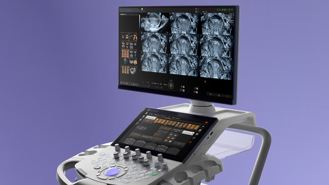
The Color Doppler Advantage
Using elite Voluson ultrasound technology, Dr. Kunda maps the baby's heart in real-time. We use "Color Doppler" to visually track the direction and speed of blood flowing through the tiny valves and chambers. This allows us to rule out holes in the heart walls, narrowed arteries, or irregular heartbeats (arrhythmias) with profound accuracy.